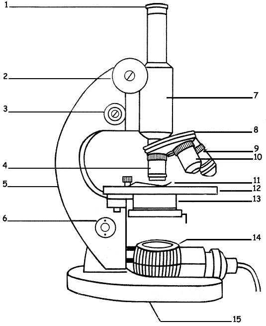
41 microscope drawing with label and function
Parts Of The Microscope Label Teaching Resources | TpT Science worksheet: Label The Parts Of A Microscope2 VERSIONS OF WORKSHEET (Worksheet with a word bank & Worksheet with no word bank)Students have to identify and label parts of microscope (Mirror, Arm, Body tube, Diaphragm, Stage, Course Focus Knob, Clip, Fine Focus Knob ,Objective Lens,Eye Piece)Second Part: Students have to match microscope parts with their functions by drawing a ... Microscope Parts, Function, & Labeled Diagram - slidingmotion Microscope parts labeled diagram gives us all the information about its parts and their position in the microscope. Microscope Parts Labeled Diagram The principle of the Microscope gives you an exact reason to use it. It works on the 3 principles. Magnification Resolving Power Numerical Aperture. Parts of Microscope Head Base Arm Eyepiece Lens
How To Draw A Microscope - YouTube Today, we're learning how to draw a cool microscope!👩🎨 JOIN OUR ART HUB MEMBERSHIP! VISIT 🎨 VISIT OUR AMAZON ART SUPPLY S...

Microscope drawing with label and function
Labeling the Parts of the Microscope | Microscope activity, Science ... Free worksheets for labeling parts of the microscope including a worksheet that is blank and one with answers. Microscope World. 113 followers. 6th Grade Science. Science Curriculum. Preschool Science. Science Classroom. Science Activities. Biology Lessons. Teaching Biology. Science Biology. Life Science ... Compound Microscope Parts, Functions, and Labeled Diagram Compound Microscope Definitions for Labels. Eyepiece (ocular lens) with or without Pointer: The part that is looked through at the top of the compound microscope. Eyepieces typically have a magnification between 5x & 30x. Monocular or Binocular Head: Structural support that holds & connects the eyepieces to the objective lenses. Parts of a Compound Microscope and Their Functions - NotesHippo Body Tube: It is the tubular structure of microscope, hollow component of the microscope arm that is attached to the top half of the arm.With the use of adjustment knobs, it can be adjusted up and down. Nose Piece: It's a spinning metal element affixed to the lower end of the body tube in a circular pattern.It has three holes for objective lenses to be inserted into.
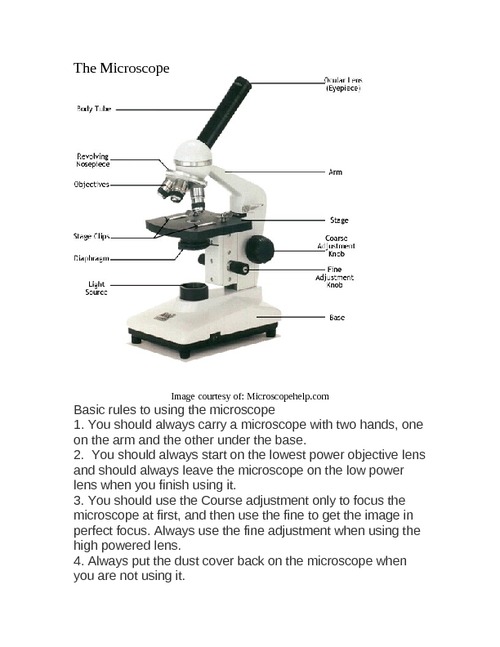
Microscope drawing with label and function. Parts of the Microscope with Labeling (also Free Printouts) 5. Knobs (fine and coarse) By adjusting the knob, you can adjust the focus of the microscope. The majority of the microscope models today have the knobs mounted on the same part of the device. Image 5: The circled parts of the microscope are the fine and coarse adjustment knobs. Picture Source: bp.blogspot.com. Microscope Drawing: How to Sketch Microscope Slides Outline the general shapes: Draw the outline of largest shape onto the paper, making it fit within the quarters. Keep you pencil drawings light and adjust the shape as needed. This may require going between the microscope slide and the drawing in order to get the proportions and shape correct. Now move to the other shapes in your field of view. Parts of a microscope with functions and labeled diagram - Microbe Notes Parts of a microscope with functions and labeled diagram April 19, 2022 by Faith Mokobi Having been constructed in the 16th Century, Microscopes have revolutionalized science with their ability to magnify small objects such as microbial cells, producing images with definitive structures that are identifiable and characterizable. Microscope Parts and Functions The specimen is placed on the glass and a cover slip is placed over the specimen. This allows the slide to be easily inserted or removed from the microscope. It also allows the specimen to be labeled, transported, and stored without damage. Stage: The flat platform where the slide is placed.
How to Sketch a Microscope Slide - Identifying and Sketching Cell ... Sketches come to life when you add highlights, shadows and color. For a pencil sketch, separate areas into white, light, medium and dark grey and black. To see the light/dark areas, squint so that the hard edges are blurred and your focus is on the shading. Start shading the light areas by following the shapes. Compound Microscope Parts - Labeled Diagram and their Functions There are two major optical lens parts of a microscope: Eyepiece (10x) and Objective lenses (4x, 10x, 40x, 100x). Total magnification power is calculated by multiplying the magnification of the eyepiece and objective lens. The illuminator provides a source of light. The light is focused by the condenser and passing through the specimen placed ... How to draw compound of Microscope easily - step by step I will show you " How to draw compound of microscope easily - step by step "Please watch carefully and try this okay.Thanks for watching.....#microscopedrawi... 18,701 Microscope drawing Images, Stock Photos & Vectors - Shutterstock Find Microscope drawing stock images in HD and millions of other royalty-free stock photos, illustrations and vectors in the Shutterstock collection. Thousands of new, high-quality pictures added every day.
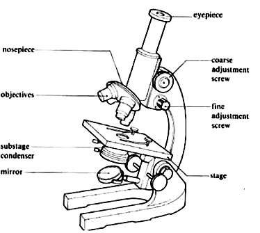
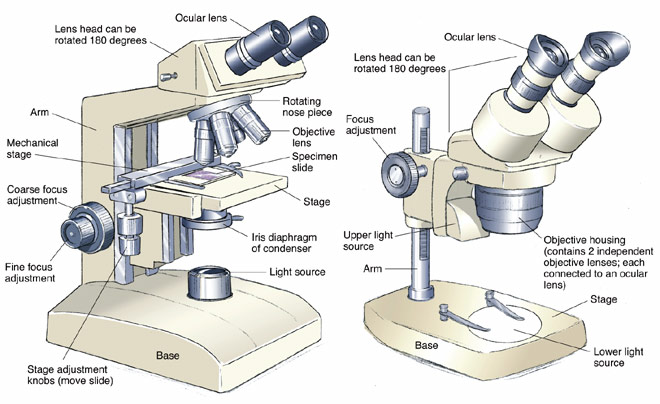
Label the microscope — Science Learning Hub All microscopes share features in common. In this interactive, you can label the different parts ... Simple Microscope - Diagram (Parts labelled), Principle, Formula and Uses Simple microscope is a magnification apparatus that uses a combination of double convex lens to form an enlarged, erect image of a specimen. The working principle of a simple microscope is that when a lens is held close to the eye, a virtual, magnified and erect image of a specimen is formed at the least possible distance from which a human eye ... Compound Microscope - Diagram (Parts labelled), Principle and Uses See: Labeled Diagram showing differences between compound and simple microscope parts Structural Components. The three structural components include. 1. Head. This is the upper part of the microscope that houses the optical parts. 2. Arm . This part connects the head with the base and provides stability to the microscope. Simple Microscope - Parts, Functions, Diagram and Labelling Stereo microscope/dissecting microscope - It can magnify objects by up to 300 times. It is used to visualize opaque objects that cannot be visualized using a compound microscope. Confocal microscope - It uses laser light to scan a dyed sample. Scanning electron microscope - Instead of light, this type of microscope uses electron. This type of microscope is used by researchers in the field of physical, biological, and medical science.
label microscope diagram | Charts | Microscope, Anatomy bones ...
Microscope, Microscope Parts, Labeled Diagram, and Functions • Step 1: Connect the light microscope to a power source in step one. You can skip this step if ...

Diagram of a Compound Microscope
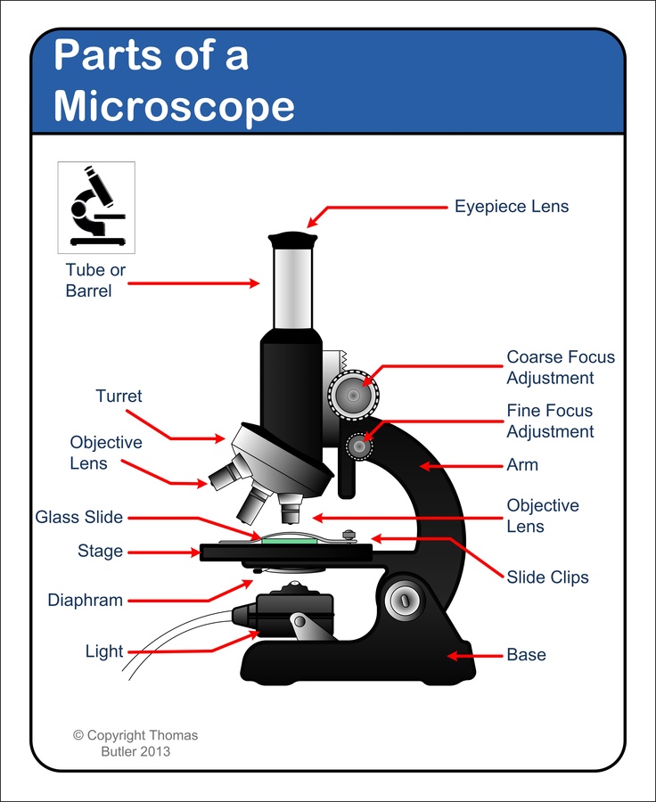
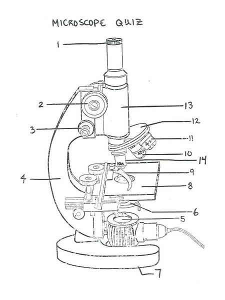
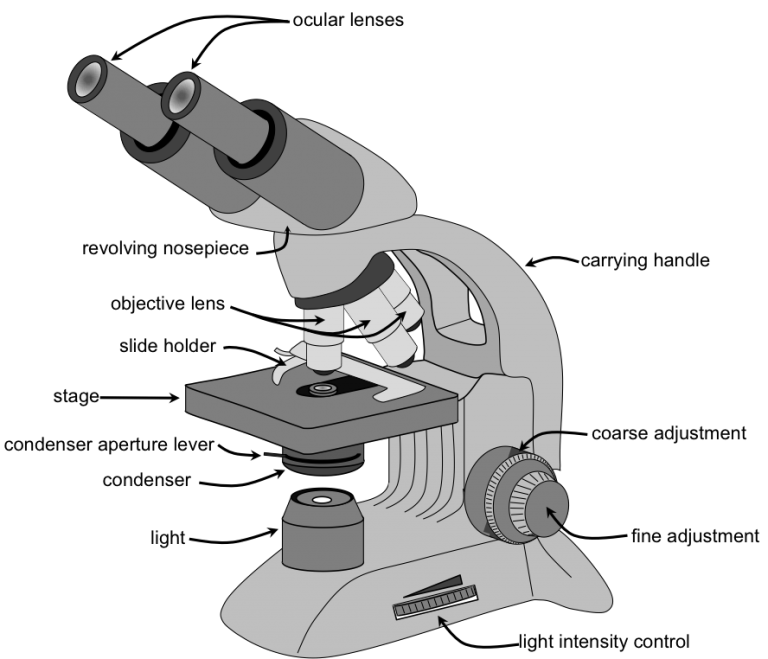
Label Microscope Diagram - EnchantedLearning.com Using the terms listed below, label the microscope diagram. arm - this attaches the eyepiece and body tube to the base. base - this supports the microscope. body tube - the tube that supports the eyepiece. coarse focus adjustment - a knob that makes large adjustments to the focus.

Compound Microscope Parts
Compound Microscope- Definition, Labeled Diagram, Principle, Parts, Uses The optical microscope often referred to as the light microscope, is a type of microscope that uses visible light and a system of lenses to magnify images of small subjects. There are two basic types of optical microscopes: Simple microscopes. Compound microscopes. The term "compound" in compound microscopes refers to the microscope having ...

Parts of a microscope with functions and labeled diagram
PDF Parts of a Microscope Printables - Homeschool Creations Label the parts of the microscope. You can use the word bank below to fill in the blanks or cut and paste the words at the bottom. Microscope Created by Jolanthe @ HomeschoolCreations.net eyepiece head objective lenses arm focusing knob base illuminator stage stage clips nosepiece.

microscope drawing | Clipart Panda - Free Clipart Images
A Study of the Microscope and its Functions With a Labeled Diagram ... A Study of the Microscope and its Functions With a Labeled Diagram To better understand the structure and function of a microscope, we need to take a look at the labeled microscope diagrams of the compound and electron microscope. These diagrams clearly explain the functioning of the microscopes along with their respective parts.

A schematic drawing of the electrical cell in combination ...
Microscope Parts & Functions - AmScope Main Microscope Parts and Functions. How to Identify Parts on a Compound Microscope. Watch on. Head: The upper part of the microscope houses the eyepiece and objective lenses. Tube: Where the eyepieces are dropped in. Also, it connects the eyepieces to the objective lenses. Stage: The flat platform that supports the slides.
Free Microscope Drawing, Download Free Microscope Drawing png ...
Parts of Stereo Microscope (Dissecting microscope) - labeled diagram ... Optical components of a stereo microscope - definition and function. Optical parts of a stereo microscope work together to magnify and produce a 3-D image of the specimens. These parts include: Eyepieces. The eyepiece (or ocular lens) is the lens part at the top of a microscope that the viewer looks through.
Simple Microscope - Parts, Functions, Diagram and Labelling ...
Labelled Diagram of Compound Microscope - Biology Discussion The below mentioned article provides a labelled diagram of compound microscope. Part # 1. The Stand: The stand is made up of a heavy foot which carries a curved inclinable limb or arm bearing the body tube. The foot is generally horse shoe-shaped structure (Fig. 2) which rests on table top or any other surface on which the microscope in kept.

Motic BA310 Digital Biological Microscope
Compound Microscope: Definition, Diagram, Parts, Uses, Working ... - BYJUS A compound microscope is defined as. A microscope with a high resolution and uses two sets of lenses providing a 2-dimensional image of the sample. The term compound refers to the usage of more than one lens in the microscope. Also, the compound microscope is one of the types of optical microscopes. The other type of optical microscope is a ...
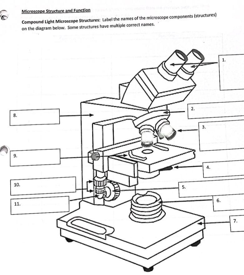
Answered: Microscope Structure and Function… | bartleby
Microscope Diagram Labeled, Unlabeled and Blank - Pinterest Microscope Diagram Labeled, Unlabeled and Blank | Parts of a Microscope. Print a microscope diagram, microscope worksheet, or practice microscope quiz in order to learn all the parts of a microscope. ... Download for free Microscope Drawing #2246314, download othes stereo microscope drawing for free. Joyliz Gonzalez. tablas. Biology Lessons ...

22 Parts Of a Microscope With Their Function And Labeled ...
Animal Cell Diagram with Label and Explanation: Cell Structure, Functions Animal cell is a typical Eukaryotic cell enclosed by a plasma membrane containing nucleus and organelles which lack cell walls, unlike all other Eukaryotic cells. The typical cell ranges in size between 1-100 micrometers. The lack of cell walls enabled the animal cells to develop a greater diversity of cell types.

Simple Microscope Definition, Magnification, Parts And Uses
Parts of a Compound Microscope and Their Functions - NotesHippo Body Tube: It is the tubular structure of microscope, hollow component of the microscope arm that is attached to the top half of the arm.With the use of adjustment knobs, it can be adjusted up and down. Nose Piece: It's a spinning metal element affixed to the lower end of the body tube in a circular pattern.It has three holes for objective lenses to be inserted into.
Free Microscope Drawing, Download Free Microscope Drawing png ...
Compound Microscope Parts, Functions, and Labeled Diagram Compound Microscope Definitions for Labels. Eyepiece (ocular lens) with or without Pointer: The part that is looked through at the top of the compound microscope. Eyepieces typically have a magnification between 5x & 30x. Monocular or Binocular Head: Structural support that holds & connects the eyepieces to the objective lenses.
Free Microscope Drawing, Download Free Microscope Drawing png ...
Labeling the Parts of the Microscope | Microscope activity, Science ... Free worksheets for labeling parts of the microscope including a worksheet that is blank and one with answers. Microscope World. 113 followers. 6th Grade Science. Science Curriculum. Preschool Science. Science Classroom. Science Activities. Biology Lessons. Teaching Biology. Science Biology. Life Science ...

Compound Microscope Drawing With Parts and Functions

13 parts of the Compound Light Microscope Diagram | Quizlet

Parts of Dissecting Microscope | Botany

Labeling the Parts of the Microscope | Microscope activity ...

List: Parts of a Microscope and their Function | Pathwooded

Microscope Parts and Functions

Parts of Stereo Microscope (Dissecting microscope) – labeled ...

microscopy how a microscope works magnification calculations ...
Microscope Parts & Function - ppt video online download

Label Microscope Diagram - EnchantedLearning.com

Simple Microscope - Diagram (Parts labelled), Principle ...
Free Microscope Drawing, Download Free Microscope Drawing png ...
Parts of a microscope with functions and labeled diagram

microscope drawing with label - Clip Art Library

Microscope Parts & Functions Part 1 (Roberts) Diagram | Quizlet

Microscope Diagram (Structures & Functions) Diagram | Quizlet
Microscope Challenge - Andrew Herrick: Teaching Portfolio

Biology 4 U on Twitter: "Try this labelled diagram Quiz on ...

Microscope Diagram and Functions | Microscope parts, Science ...

Laboratory application of imaging technology on pavement ...
Compound Microscope - Types, Parts, Diagram, Functions and ...
Free Microscope Drawing, Download Free Microscope Drawing png ...
22 Parts Of a Microscope With Their Function And Labeled ...

Compound Microscope: Parts of Compound Microscope

SOLUTION: Compound microscope parts and functions - Studypool
مجهر بيولوجي
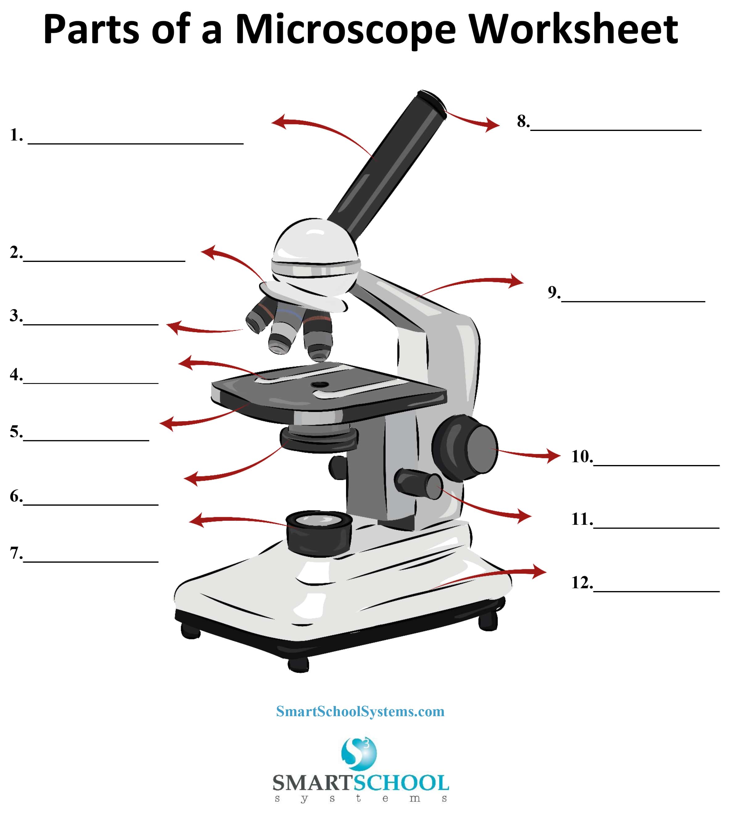
Parts of a Microscope - SmartSchool Systems
Post a Comment for "41 microscope drawing with label and function"