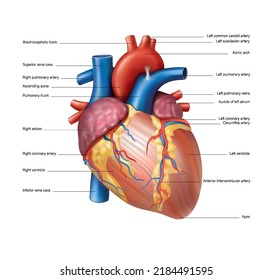
42 anterior view of the heart with labels
anterior view of heart region of cadaver Diagram | Quizlet anterior view of heart region of cadaver Diagram | Quizlet anterior view of heart region of cadaver STUDY Learn Write Test PLAY Match + − Created by roconnor728 Terms in this set (18) brachiocephalic trunk ... brachycephalic veins ... superior vena cava ... right lung ... trachea ... left common carotid artery ... left subclavian artery ... Coronary artery dominance - wikidoc When present, it is one of the three longest branches on the inferior wall of the heart. 10: RP: Right posterior artery: Distal most branch to arise from the right coronary artery, but present only in large right dominant systems. When present, it is one of the three longest branches on the inferior wall of the heart. 26: C4: Left ...
Heart - Collection Page | AnatomyTOOL 3D model of the normal heart and lungs with numbered English labels. From a collaboration of Universities of Leiden, Delft and Groningen. ... A photo of the anterior view of a plastinated normal heart, where highlights on structures can be switched on and off, by Univ. of Br. Columbia.

Anterior view of the heart with labels
Heart chambers and associated great vessels - Anatomy Image: Anterior view of the human heart with labels. right atrium has two basic parts: a smooth posterior and an anterior portion in which bundles of muscle tissue form ridges in the walls. The muscle bundles are called pectinate muscles because they look like the teeth of a comb. anterior heart Quiz - PurposeGames.com This is an online quiz called anterior heart There is a printable worksheet available for download here so you can take the quiz with pen and paper. From the quiz author quiz game to assist a&p students with anatomy of exterior heart. This quiz has tags. Click on the tags below to find other quizzes on the same subject. quiz heart label anterior Solved Label the structures seen in the anterior view of the - Chegg View the full answer Transcribed image text: Label the structures seen in the anterior view of the heart. Superior vena cava Interior vena cava Aorta Left atrium Pulmonary trunk Pulmonary vein Left ventricle Pulmonary artery Right ventricle Right atrium Previous question Next question
Anterior view of the heart with labels. Anatomy of the foot and ankle - MRI - e-Anatomy - IMAIOS Aug 26, 2022 · By moving the mouse cursor over a specific area, this region is highlighted and its labels displayed: anterior and posterior region of the leg, posterior and anterior ankle region, metatarsal region, plantar region. The vertical menu on the left provides cross-references and a 3D medical illustration of the foot skeleton. Male Human Anatomy Diagram Pictures, Images and Stock Photos The human body and a skeleton with a silhouette of a body. A male, female person standing. Front view, side view in full length. Adult and kid x-ray image. People anatomy. A vector illustration on a white background. male human anatomy diagram stock illustrations Whitesnake (album) - Wikipedia Initially the album was released worldwide with different titles, tracklists and by different record labels. In Europe and Australia, it was titled 1987 and included two extra songs absent from the North American version, "Looking for Love" and "You're Gonna Break My Heart Again", while in Japan the album was released as Serpens Albus with the ... Lab 44- Heart Structure Flashcards | Quizlet Label the anterior heart structures by clicking and dragging the labels to the correct location. 1. Aoric Arch 2.Super vena cava 3. Righ pulmonary arteries 4. Ascending aorta 5. Right pulmonary veins 6.Right auricle 7.Right coronary artery. 8.Small cardiac vein 9.Marginal artery 10.Right ventricle 11. Inferior vena Cava (Straight down.)
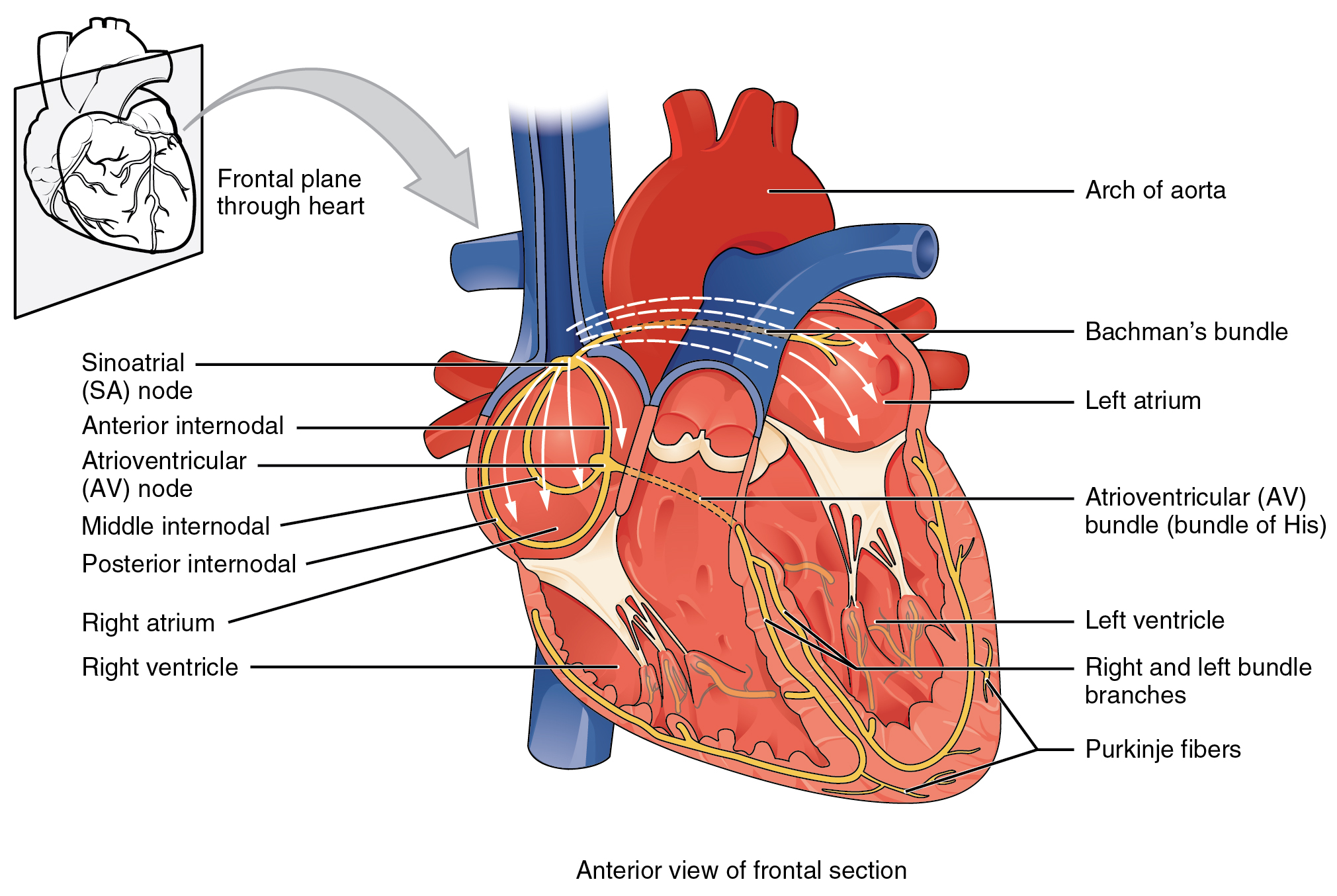
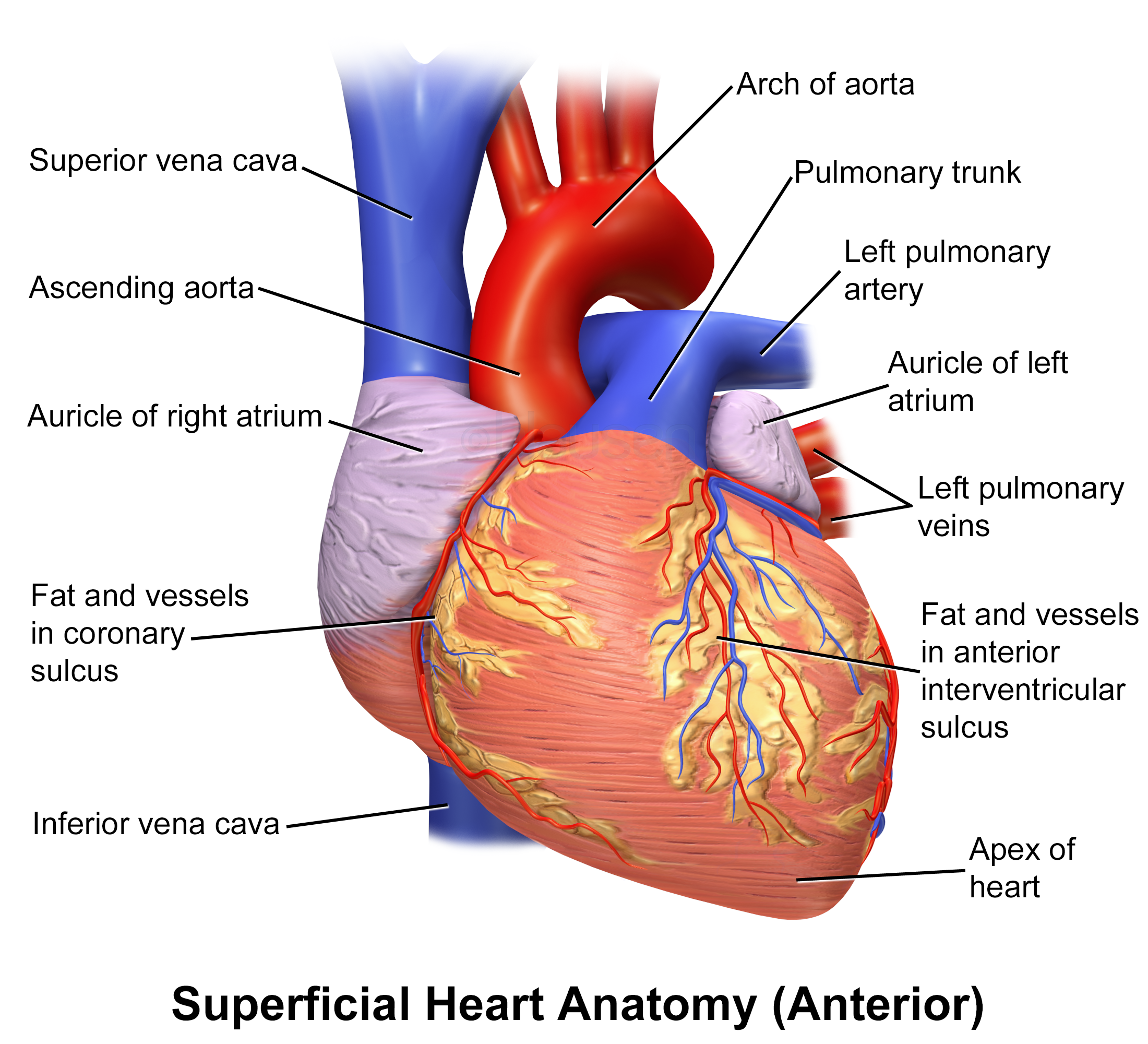
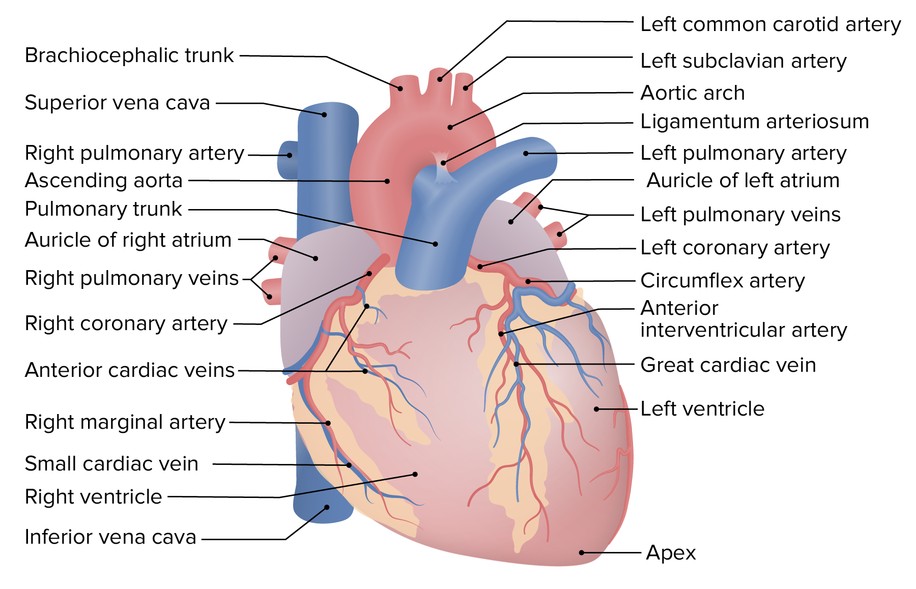
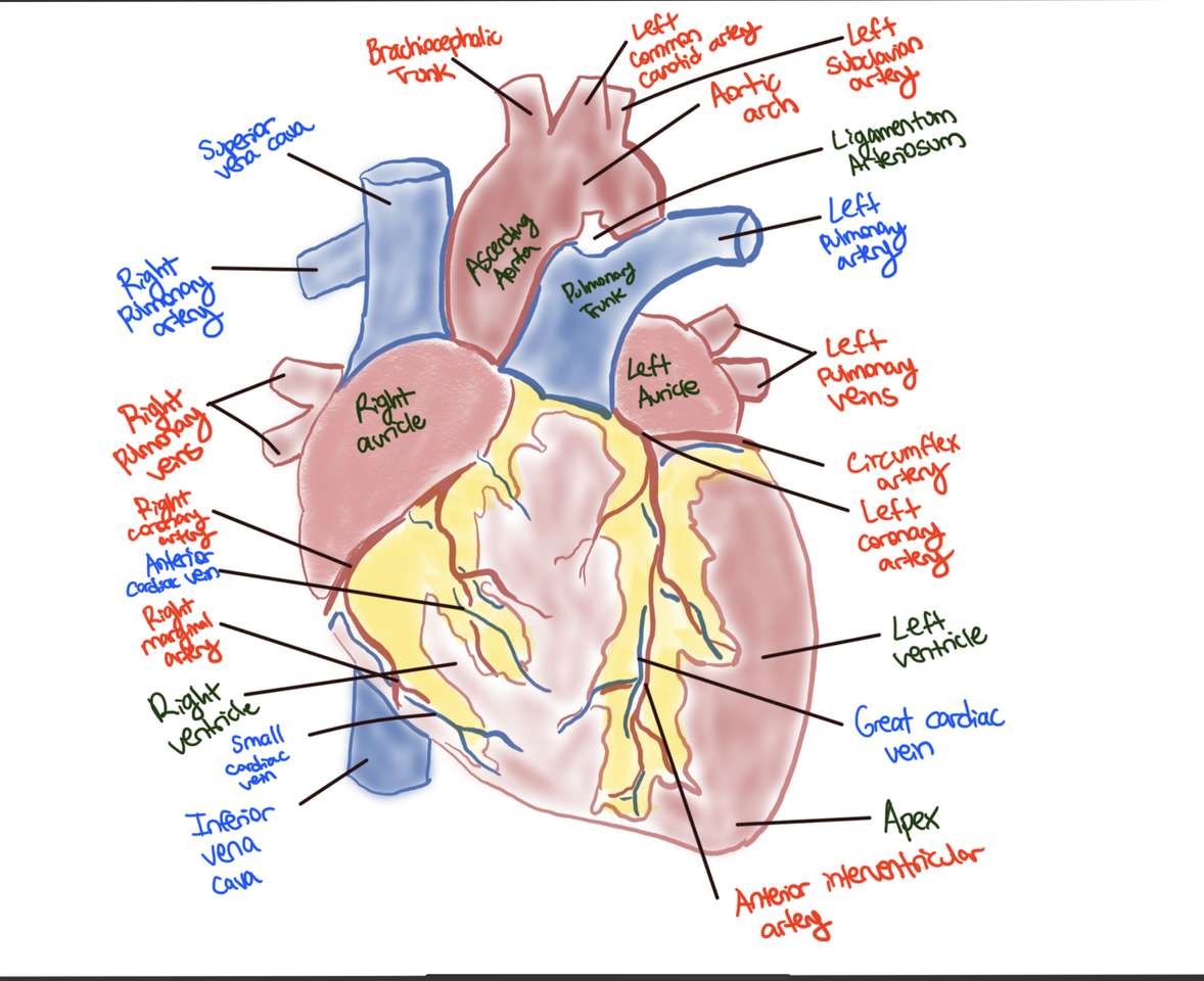
Label the Heart Diagram Anterior view - Printable - PurposeGames.com About this Worksheet This is a free printable worksheet in PDF format and holds a printable version of the quiz Label the Heart Diagram Anterior view. By printing out this quiz and taking it with pen and paper creates for a good variation to only playing it online. Figure 2.1: Anatomy of the Heart (Anterior View): The figure... Atrial fibrillation (AF) is the most common cardiac arrhythmia in United States. The most popular treatment for AF is a percutaneous procedure called catheter ablation. Current AF ablation... Anatomy Tutorial - Anterior | Atlas of Human Cardiac Anatomy This illustration demonstrates an anterior view of the thoracic cavity, highlighting the position of the heart in relationship to the ribs and diaphragm. The right atrium, right ventricle, and a small portion of the left ventricle are visible from this aspect. Note that in the majority of cases, 2/3 of the heart is positioned to the left of ... Chapter 19: The Heart Flashcards | Quizlet -Heart muscle receives blood when the ventricles relax •Left coronary artery (LCA)-anterior interventricular branch •supplies blood to interventricular septum and anterior walls of ventricles-circumflex branch •passes around left side of heart in coronary sulcus, supplies left atrium and posterior wall of left ventricle
Label the Heart Diagram Anterior view Quiz - PurposeGames.com This is an online quiz called Label the Heart Diagram Anterior view There is a printable worksheet available for download here so you can take the quiz with pen and paper. Your Skills & Rank Total Points 0 Get started! Today's Rank -- 0 Today 's Points One of us! Game Points 24 You need to get 100% to score the 24 points available Actions 2 favs Jain - Drawing Anterior view of heart - no labels | AnatomyTOOL Anterior view of heart. The heart and its great vessels are drawn in this image, also the coronary arteries and veins are shown. This image was retrieved from OERCommons.org Anatomical structures in item: Cor Aorta Arcus aortae Aorta ascendens Vena cava superior Vena cava inferior Atrium dextrum Atrium sinistrum Arteria coronaria sinistra Sheep Heart - San Diego Mesa College SHEEP HEARTS. Click on a photo for a larger view of the model. Click on Label for the labeled model. Back to Dissected Specimen Page ch. 13 Homework Flashcards | Quizlet Label the structures seen in an anterior view of the heart. This flow diagram illustrates the effects of an increased cardiac output and the role of baroreceptors in controlling changes in blood pressure. Place the labels in the correct location based on the hints provided. 1. Cardiac output increases 2. blood pressure rises
Lab Prep: Thorax and Heart Flashcards | Quizlet
Blausen 0451 - Anterior view of the heart - English labels Reviewed Information Download. 814 reads. Blausen 0451 - Anterior view of the heart - English labels. Uploaded by: Student10. Institution: Netherlands, Leiden - Leiden University Medical Center, Leiden University. Creator (s)/credit: Blausen Medical Communications, Inc. 9/10.
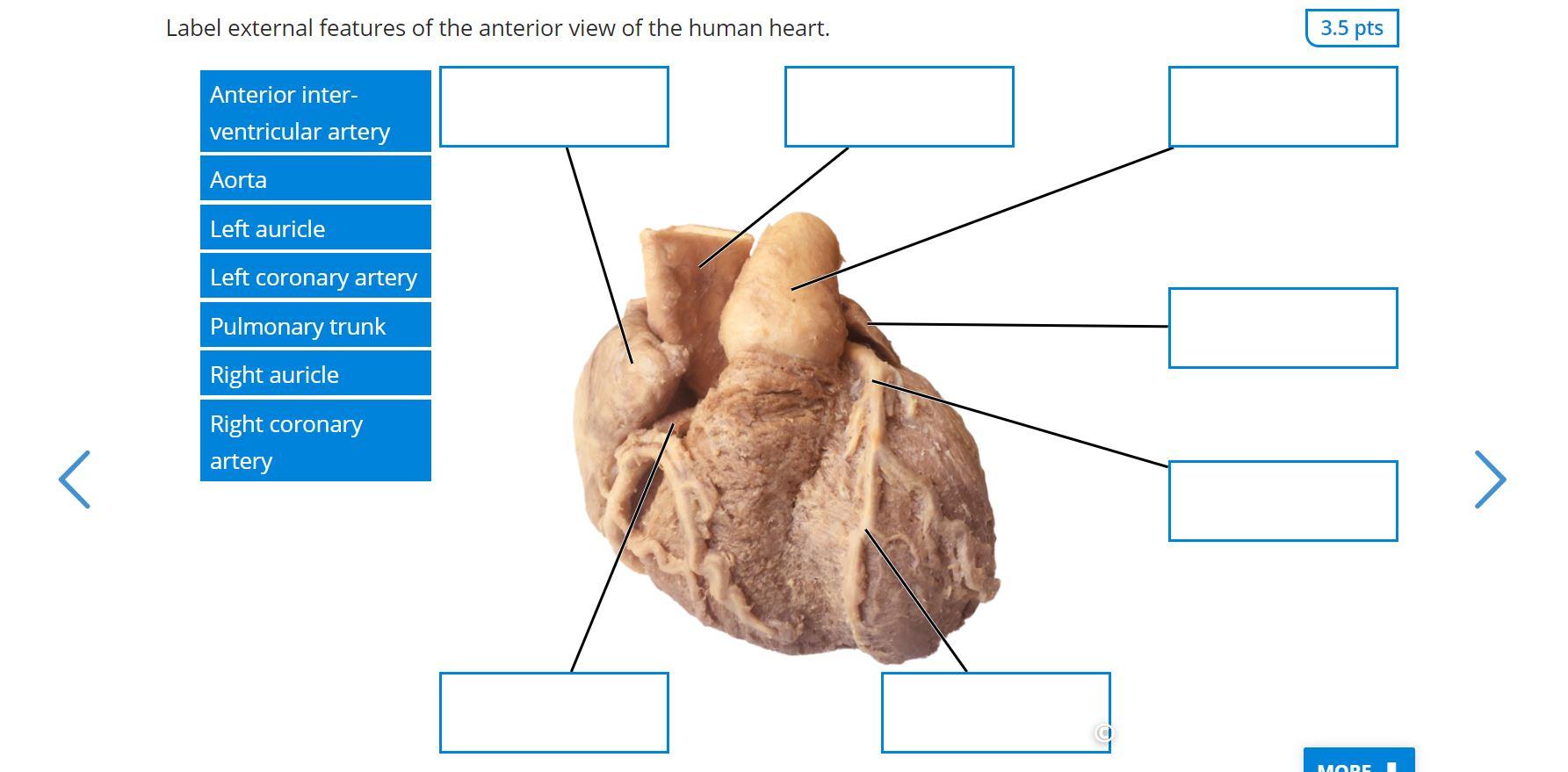
Solved Label external features of the anterior view of the ...
Norepinephrine - Wikipedia Norepinephrine (NE), also called noradrenaline (NA) or noradrenalin, is an organic chemical in the catecholamine family that functions in the brain and body as both a hormone and neurotransmitter.
File:2018 Conduction System of Heart.jpg - Wikimedia Commons
External Anterior View of Heart Quiz - PurposeGames.com Latest Activities. An unregistered player played the game 1 week ago; An unregistered player played the game 2 weeks ago; An unregistered player played the game 2 ...
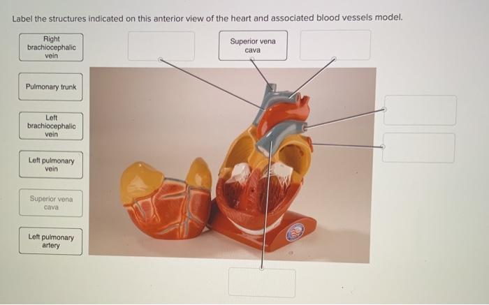
Solved Label the structures indicated on this anterior view ...
147 Heart Anatomy With Labels Premium High Res Photos - Getty Images Browse 147 heart anatomy with labels stock photos and images available, or start a new search to explore more stock photos and images. of 3. NEXT.

Heart Lab Flashcards | Quizlet
Heart bypass surgery - minimally invasive - MedlinePlus Your doctor may recommend a minimally invasive coronary artery bypass if you have a blockage in one or two coronary arteries, most often in the front of the heart. When one or more of the coronary arteries become partly or totally blocked, your heart does not get enough blood. This is called ischemic heart disease or coronary artery disease.

Heart and great vessels (anterior view). The heart is ...
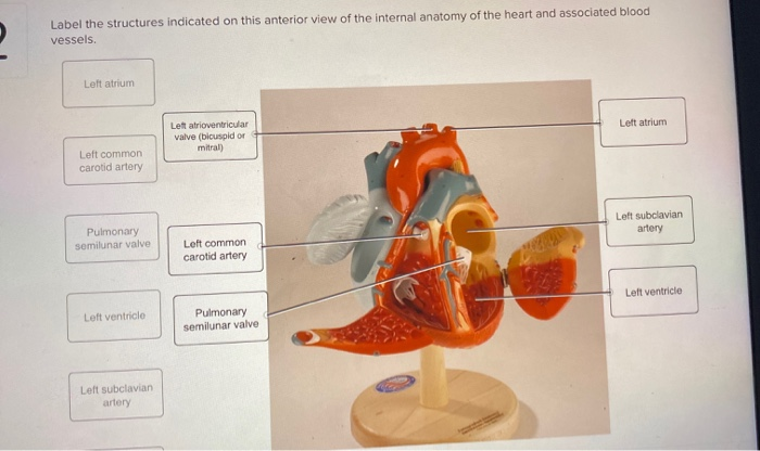
Solved Label the structures indicated on this anterior view - Chegg Expert Answer 100% (7 ratings) Markings above the figure 1. Righ … View the full answer Transcribed image text: Label the structures indicated on this anterior view of the internal anatomy of the heart model. Left Atrium Interventricular septum Chordae tendineae Left AV Valve (bicuspid or mitral) Right atrium Right AV Valve (tricuspid)

BIO 114 - Anterior View of the Heart Quiz - By tgardiner9
Label the heart — Science Learning Hub In this interactive, you can label parts of the human heart. Drag and drop the text labels onto the boxes next to the diagram. Selecting or hovering over a box will highlight each area in the diagram. pulmonary vein. semilunar valve. right ventricle. right atrium. vena cava. left atrium.

anterior view of the heart Diagram | Quizlet
Heart Anatomy | Anatomy and Physiology II - Lumen Learning The dorsal surface of the heart lies near the bodies of the vertebrae, and its anterior surface sits deep to the sternum and costal cartilages. The great veins, the superior and inferior venae cavae, and the great arteries, the aorta and pulmonary trunk, are attached to the superior surface of the heart, called the base.

Heart anatomy anterior view without labeled
Heart Diagram with Labels and Detailed Explanation - BYJUS Diagram of Heart. The human heart is the most crucial organ of the human body. It pumps blood from the heart to different parts of the body and back to the heart. The most common heart attack symptoms or warning signs are chest pain, breathlessness, nausea, sweating etc. The diagram of heart is beneficial for Class 10 and 12 and is frequently ...
Solved] Label the structures indicated on this anterior view ...
Chapter 22 Heart Flashcards | Quizlet Label the coronary arteries in an anterior view of the heart. Label the order that blood flows through in the heart, using the arrows as guides. Label the components of the heart wall. Label the components of the heart as seen from a posterior view. Label the major coronary veins. Label the components of the conduction system.

1 shows the heart anatomy from the anterior and interior ...
Solved Heart Chambers and Valves: Frontal Heart Section 2 - Chegg Heart Chambers and Valves: Frontal Heart Section 2 Label the chambers and valves seen in an anterior view of the heart Left ventricle Right ventricle Aortic valve Tricuspid Valve Right atrum Mihral valve Chord Pulmonary ave
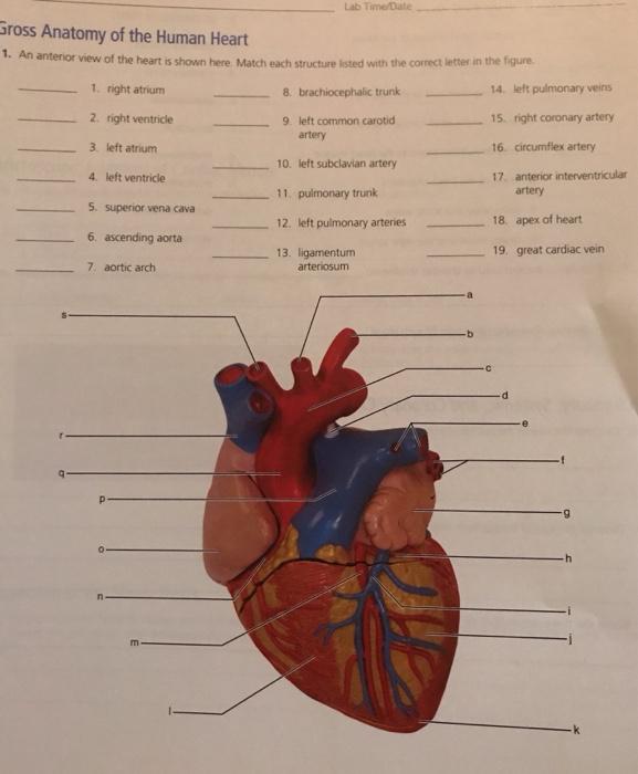
Solved Lab Time/Date Gross Anatomy of the Human Heart 1. An ...
Lab heart label 1.pdf - Anterior View of Heart Label:... View Lab heart label 1.pdf from BIO 103B at Ohlone College. Anterior View of Heart Label: Brachiocephalic Trunk Left Common Carotid artery Aortic arch

External Heart Anatomy Quiz
Internal view of the heart label yourself.png - | Course Hero External view of the heart label yourself.png. 1. Anterior cross-section view of heart.png. 1. Anterior view-anatomy of heart.png. 1. Arteries of the head and neck.png. 1. Arteries of the abdomen and pelvis.png.

Chart of heart anterior view with parts name - vector image ...
Heart Anatomy: Labeled Diagram, Structures, Blood Flow ... - EZmed There are 4 chambers, labeled 1-4 on the diagram below. To help simplify things, we can convert the heart into a square. We will then divide that square into 4 different boxes which will represent the 4 chambers of the heart. The boxes are numbered to correlate with the labeled chambers on the cartoon diagram. View fullsize
Anterior (frontal) view of the opened heart [5]. | Download ...
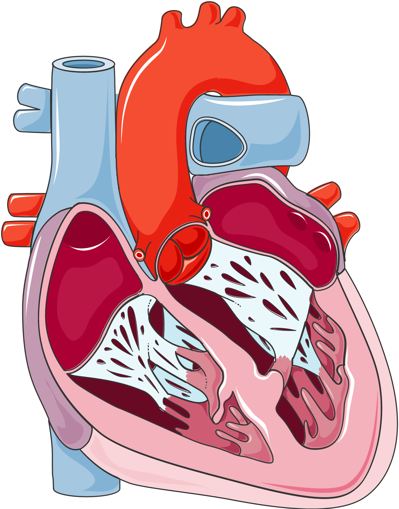
The following diagram depicts the interior of the heart anterior view ... 4. The following diagram depicts the interior of the heart (anterior view). Label the parts indicated. 5. a) The papillary muscles are columns of myocardium that project into each ventricle (chamber) of the heart. b) The chordae tendineae are strands of fibrous connective tissue that extend from the papillary muscles to the Flaps of the AV VALVES c) The function of these structures is to ...

Human Heart Anterior View High-Res Vector Graphic - Getty Images
Heart Labeling anterior view Diagram | Quizlet Heart Labeling anterior view STUDY Learn Write Test PLAY Match + − Created by Meghan12th PLUS Terms in this set (26) brachiocephalic trunk ... left common carotid artery ... superior vena cava ... aortic arch ... liigamentum arteriosum ... right pulmonary artery ... amending aorta ... right pulmonary veins ... pulmonary trunk ... right atrium ...

Pin on Paramedic Study Guide
Heart Anatomy - Anterior (Front) View : Medical Illustration Item ID: si55550728 Source ID: 2. Request Pricing. Description: This medical exhibit pictures an anterior (front) view of the heart anatomy with labels for the aorta, superior vena cava, right atrium, right ventricle, inferior vena cava, pulmonary trunk, left atrium, pulmonary veins and left ventricle. Max Image Size: 2448 pixels wide by 1804 ...

heart diagram - Google Search | Heart structure, Human body ...
Solved Label the structures seen in the anterior view of the - Chegg View the full answer Transcribed image text: Label the structures seen in the anterior view of the heart. Superior vena cava Interior vena cava Aorta Left atrium Pulmonary trunk Pulmonary vein Left ventricle Pulmonary artery Right ventricle Right atrium Previous question Next question
Servier - Drawing Coronal section of the heart anterior view ...
anterior heart Quiz - PurposeGames.com This is an online quiz called anterior heart There is a printable worksheet available for download here so you can take the quiz with pen and paper. From the quiz author quiz game to assist a&p students with anatomy of exterior heart. This quiz has tags. Click on the tags below to find other quizzes on the same subject. quiz heart label anterior
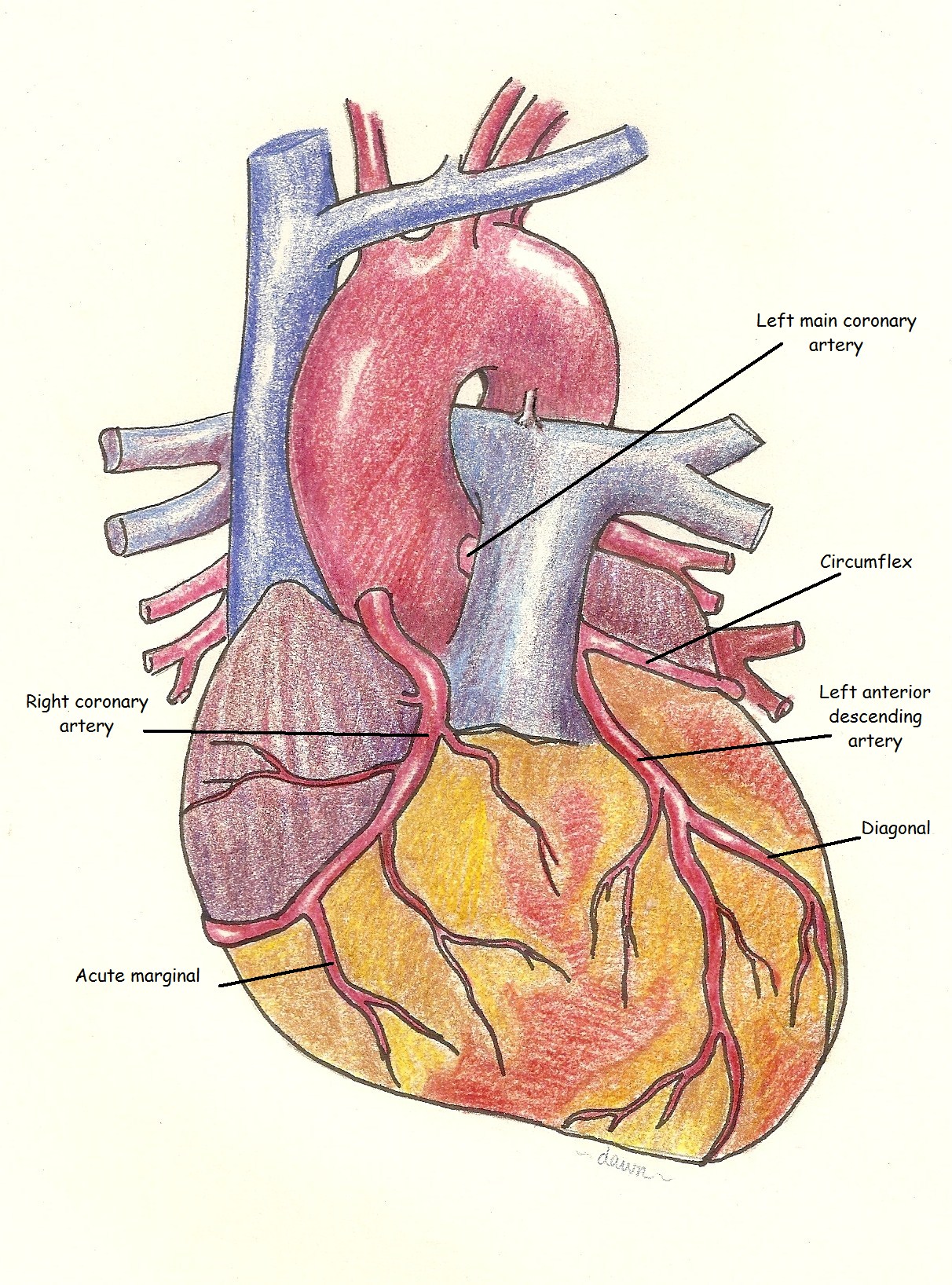
Coronary Arteries Anterior View Labeled | ECG Guru ...
Heart chambers and associated great vessels - Anatomy Image: Anterior view of the human heart with labels. right atrium has two basic parts: a smooth posterior and an anterior portion in which bundles of muscle tissue form ridges in the walls. The muscle bundles are called pectinate muscles because they look like the teeth of a comb.

Cardiovascular System | Human Anatomy | Life Science ...

1: Anatomy of the Heart (Anterior View): The figure ...
What are the parts of the heart? - Quora

anterior heart Quiz

Heart Anatomy Lab Quiz Anterior View Diagram | Quizlet
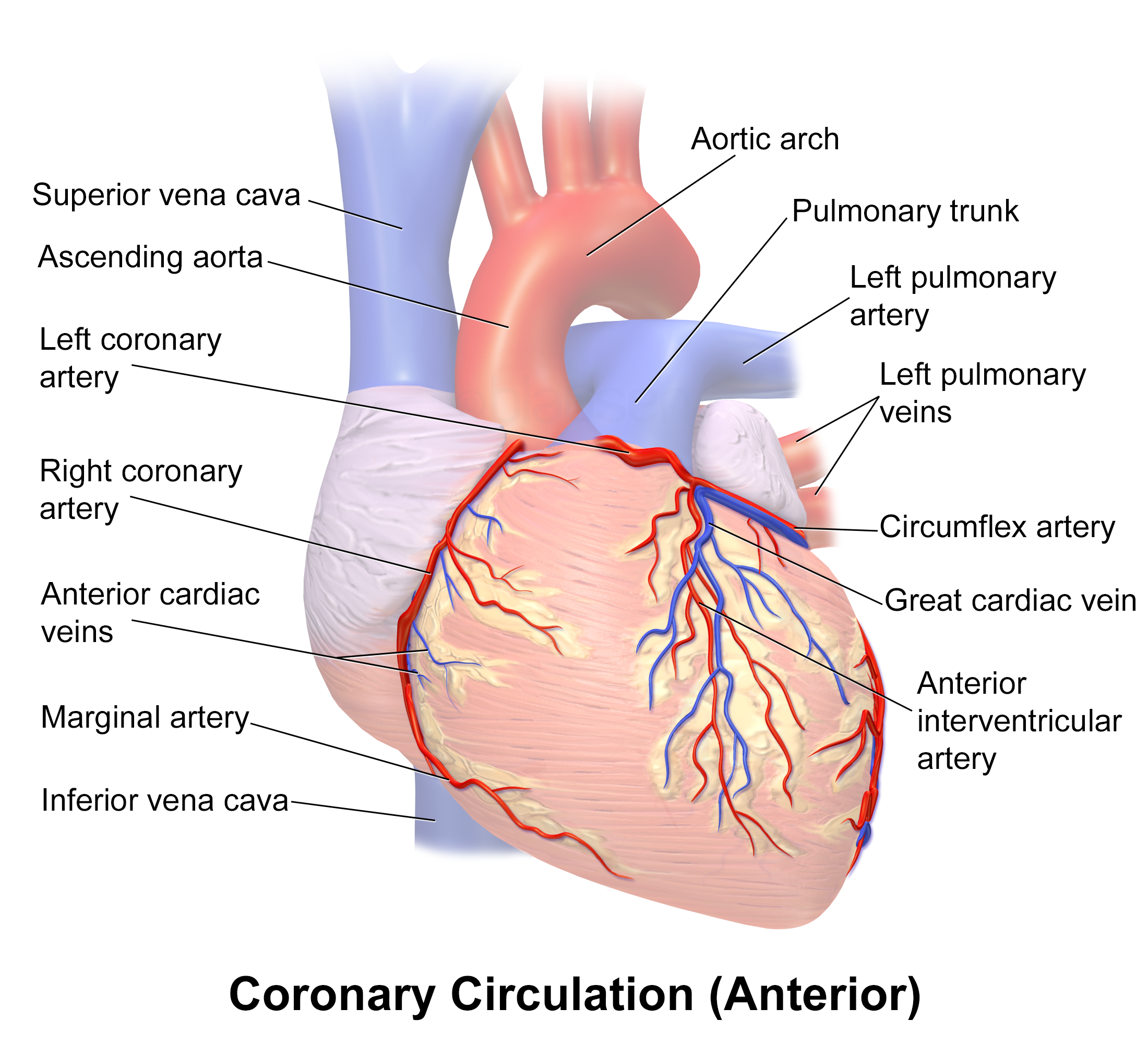
Blausen 0260 - Coronary vessels (Anterior view) - English ...
648 Human heart 3d with label Images, Stock Photos & Vectors ...
Blausen 0451 - Anterior view of the heart - English labels ...

An anterior view of the heart... | Download Scientific Diagram

Anterior View of the Human Heart (preview) - Human Anatomy | Kenhub
Solved Label the structures indicated on this anterior view ...
What are the parts of the heart? - Quora
Heart: Anatomy | Concise Medical Knowledge

Cardiovascular (A&P) CH. 15 Flashcards | Quizlet
Anatomy of heart anterior view - ePuzzle photo puzzle
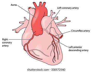
Coronary Arteries Heart Anterior View Including Stock Vector ...

Label With Me - Anatomy of the Heart (Anterior View) - YouTube

Heart Anatomy: Labeled Diagram, Structures, Blood Flow ...

External anterior view of the heart contd Diagram | Quizlet

ORGIL ORGIO (orgiodr) - Profile | Pinterest
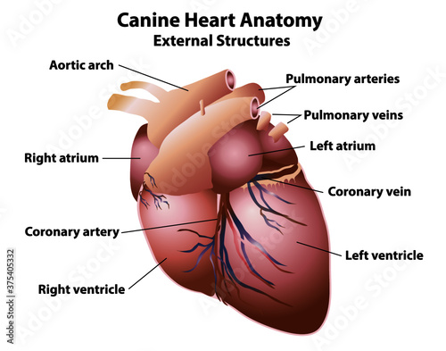
Canine heart with anatomy labels. External dog heart ...
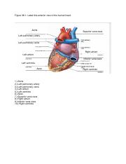
Lab Report 38 Figures 38.1, 38.2, and 38.3.pdf - Figure 38.1 ...
Post a Comment for "42 anterior view of the heart with labels"