41 skin label diagram
Label the diagram of the skin - Labelled diagram - Wordwall Label the diagram of the skin - Labelled diagram Hair shaft, Hair follicle, Arrector pili muscle, Sweat pore, Sebaceous gland, Sweat gland, Blood capillaries, Dermal Papila, Nerve endings, Epidermis, Dermis, Subcutaneous layer. Label the diagram of the skin Share by Charlotte98 Biology Like Edit Content More Log in required Theme Log in required Label the Skin Quiz - PurposeGames.com This is an online quiz called Label the Skin. There is a printable worksheet available for download here so you can take the quiz with pen and paper. Your Skills & Rank. Total Points. 0. Get started! Today's Rank--0. Today 's Points. One of us! Game Points. 11. You need to get 100% to score the 11 points available.
Anatomy, Diagram and Function of Skin - VEDANTU The skin has a surface area of between 16.1-21.5 sq ft. for an adult human. The thickness of the skin differs over all parts of the body, and between men and women and the young and the old. For example, the skin on the forearm which is on average 1.3 mm in the human male and 1.26 mm in the human female.

Skin label diagram
Integumentary system parts: Quizzes and diagrams | Kenhub Spend some time analyzing the skin diagram labeled above. Try to memorize the appearance and location of each structure. Learning the function of each structure will accelerate your ability to memorize, so be sure to check out our detailed article on The Integumentary System parts and functions. ... skin labeling Diagram | Quizlet Epidermis. outermost layer of the skin;composed of squamous epithelium; contains keratin. subcutaneous layer. Innermost layer of skin, contains fat and is the location of main blood vessels. basal layer of epidermis. The deepest layer of the Epidermis (outermost layer of the skin). The cells in the basal layer are alive, multiplying and growing. Skin Labeling | Biology Game | Turtle Diary Identify and label figures in Turtle Diary's interactive online game, Skin Labeling! Drag the given words to the correct blanks to complete the labeling! Skin Labeling - Biology Game. Bly8337. 35,274 Plays Grade K - 5 (650) Skin Labeling.
Skin label diagram. Skin Diagram || How to draw and label the parts of skin - YouTube 'Skin Diagram || How to draw and label the parts of skin' is demonstrated in this video tutorial step by step.The sense of touch had received supreme importa... Layers of Skin: How Many, Diagram, Model, Anatomy, In Order Layers of Skin: How Many, Diagram, Model, Anatomy, In Order The Layers of Your Skin Your skin is your body's largest external organ. It provides a barrier between your body's essential organs,... Skin 1: the structure and functions of the skin - Nursing Times Nurses need to understand the skin and its functions to identify and manage skin problems. This article, the first in a two-part series, looks at the skin's structure and key functions. This article comes with a self-assessment enabling you to test your knowledge after reading it. Abstract. Skin diseases affect 20-33% of the population at any ... Skin Diagram Worksheets | 99Worksheets Skin Diagram Worksheets. The Anatomy And Physiology Of Animalsskin Worksheetworksheet. Integumentary System Diagram To Label Awesome Free Coloring Pages. Skin Worksheet. Skin Diagram Worksheet Have Fun Teaching. Free Skin Diagram printable Science worksheets for 5th Grade students. Click on the image to view or download the PDF version.
Anatomy, Skin (Integument), Epidermis - StatPearls - NCBI Bookshelf Stratum lucidum, 2-3 cell layers, present in thicker skin found in the palms and soles, is a thin clear layer consisting of eleidin which is a transformation product of keratohyalin. Stratum corneum, 20-30 cell layers, is the uppermost layer, made up of keratin and horny scales made up of dead keratinocytes, known as anucleate squamous cells. The Skin (Human Anatomy): Picture, Definition, Function, and Skin ... The skin is the largest organ of the body, with a total area of about 20 square feet. The skin protects us from microbes and the elements, helps regulate body temperature, and permits the ... A Human Body Skin-structure Quiz! - ProProfs Quiz Flashcard. Create your own Quiz. In this, a human body skin structure quiz, we are going to focus on the underlying and the most elementary structure of the human body. It's easy to take your skin for granted, but when you consider how it protects your body from harm, it is something we should appreciate more. Skin Anatomy - EnchantedLearning.com Human skin is only about 0.07 inches (2 mm) thick. Skin is made up of two layers that cover a third fatty layer. The outer layer is called the epidermis; it is a tough protective layer that contains melanin (which protects against the rays of the sun and gives the skin its color).
Label Skin Diagram Printout - EnchantedLearning.com Read the definitions, then label the skin anatomy diagram below. blood vessels - Tubes that carry blood as it circulates. Arteries bring oxygenated blood from the heart and lungs; veins return oxygen-depleted blood back to the heart and lungs. dermis - (also called the cutis) the layer of the skin just beneath the epidermis. Skin diagram labeled - Healthiack This brief article displays Skin diagram labeled … Please click on the diagram (s) to view larger version. You are welcome to browse healthiack.com for more details on this very topic. Best viewed on 1280 x 768 px resolution in any modern browser. Skin diagram labeled 1075 Skin diagram labeled 1077 Skin diagram labeled 1080 Labeled Skin Structure Diagram | Quizlet Labeled Skin Structure STUDY Learn Flashcards Write Spell Test PLAY Match Gravity Created by Styson64TEACHER Figure 4.3 from pg 114 of your textbook Terms in this set (20) skin structure Hair Shaft Nonliving, extracellular matrix produced and secreted by hair follicle cells. Involved in protection, sensation, and temperature regulation Epidermis Pin by Benjamin Brown on Pathogenicity | Skin anatomy, Integumentary ... This article will look at the components and the accessory structures of the integumentary system, skin healing, skin integrity, and the staging of pressures ulcers. This article contains 7 Facts about the Integumentary System Every Nursing Student Should Know. #nursecepts #iintegumentarysystem #nursingstudent #nursingschool. N.
Illustration Of Blistering Second Degree Burn In Epidermis And Dermis ...
Skin Diagram To Label Illustrations, Royalty-Free Vector Graphics ... Vector butcher block skin diagram to label stock illustrations. Hair graft types set for hair transplantation Hair micrograft classification set for hair transplantation surgery. Skin cross-section with number of hairs in the follicular unit or family. Hair science and anatomy. Cartoon vector illustration. skin diagram to label stock illustrations
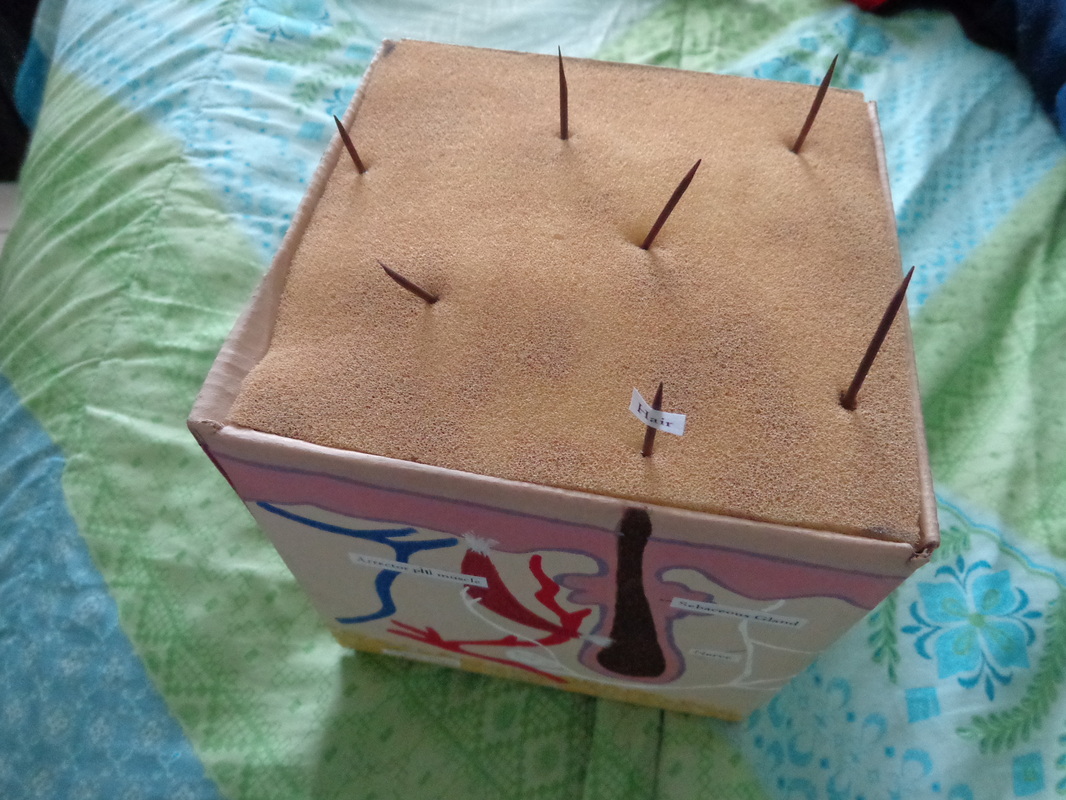
Anatomy 3D Skin Model - Megan Hammer
Skin Diagram with Detailed Illustrations and Clear Labels Important Diagrams Skin Diagram The largest organ in the human body is the skin, covering a total area of about 1.8 square meters. The skin is tasked with protecting our body from the external elements as well as microbes. Interesting Note:
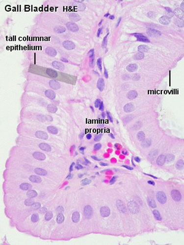
ANAT2241 Liver, Gallbladder, and Pancreas - Embryology
Skin Histology Slide Identification - AnatomyLearner Skin histology slide labeled diagram Again, thin skin is found on the ventral surface of the body and the medial surface of the limb. Thin skin has a protective hair coat. In the histology slide, you will find the thinner epidermis in thin skin. Let's identify the thick and thin skin histology slides under a light microscope.
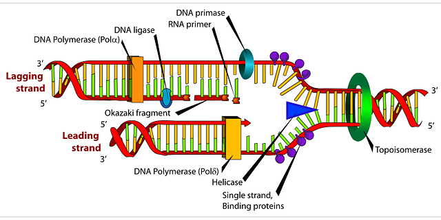
Diagram Dna Biology · Free vector graphic on Pixabay
Skin Labeling Quiz - PurposeGames.com This is an online quiz called Skin Labeling. There is a printable worksheet available for download here so you can take the quiz with pen and paper. From the quiz author. Epidermis, Dermis, Hypodermis Your Skills & Rank. Total Points. 0. Get started! Today's Rank--0. Today 's Points. One of us!

Annelida roundworm diagram | Earthworms, Worms, Diagram
Skin diagram to label - Labelled diagram - Wordwall Skin diagram to label - Labelled diagram Epidermis, Dermis, Hypodermis, Blood and lymph, Sensory nerve ending, Sweat gland, Arrector pili muscle, Sebaceous gland, Hair shaft, Dermal papilla, Hair follicle. Skin diagram to label Share by Kmiller14 Like Edit Content More Leaderboard Log in required Theme Log in required Options Switch template
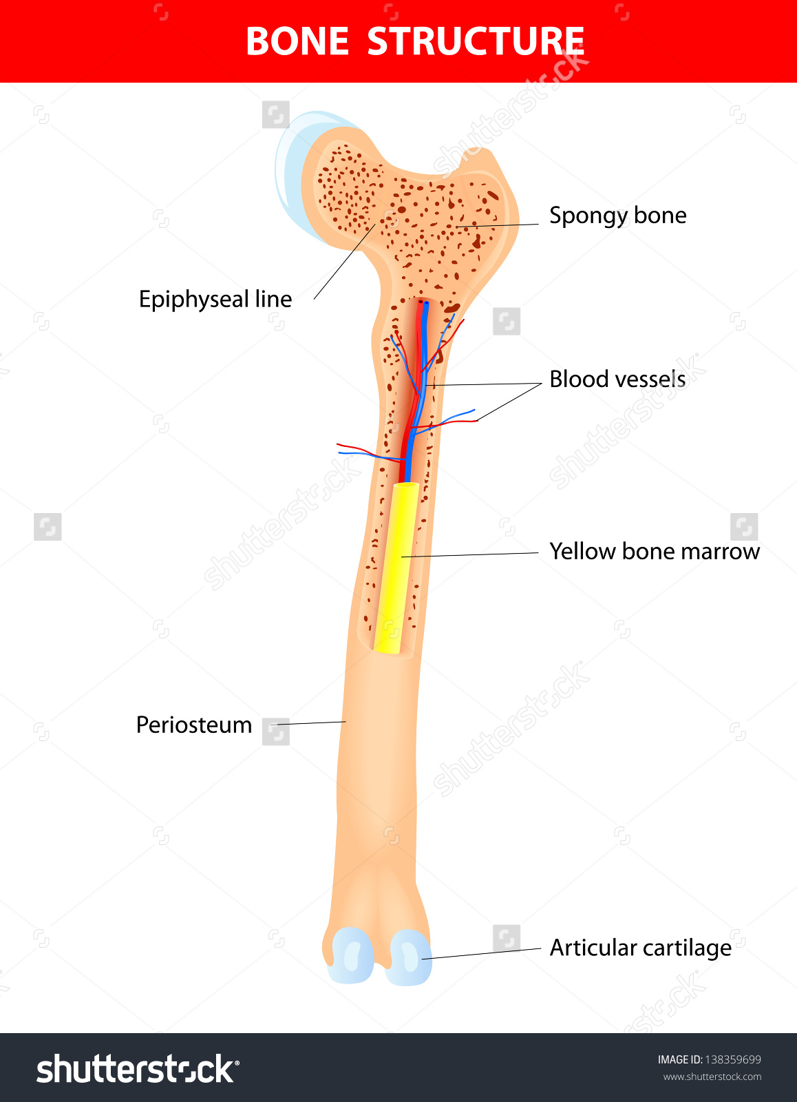
Bone structure clipart - Clipground
Skin Worksheet Answers - WikiEducator Skin Worksheet Answers. 1. The outermost layer of the skin is: the epidermis. 2. Which is the thickest layer: the dermis. 3. Add labels to the diagram of the skin shown below. 4. Which of the following happens to epidermal cells as they move up to the surface of the skin?

Lamellar corpuscle - YouTube
PDF Skin Diagram Labeling - New Providence School District Skin Diagram Labeling . 1. Label the diagram with the . letters. below according to the structure/area they describe. You may label with a line or put the label directly onto the area described. Be as precise as possible. If you are worried about the precision of your label add a word after to explain exactly where your label should be.
Elements of Morphology: Human Malformation Terminology
Label_the_Skin_Anatomy_Diagram_AP_online.docx - Name_ Date _ Period ... Name_____ Date _____ Period_____ Label the Skin Anatomy Diagram Read the definitions, then label the skin anatomy diagram below. blood vessels - Tubes that carry blood as it circulates. Arteries bring oxygenated blood from the heart and lungs; veins return oxygen-depleted blood back to the heart and lungs. dermis - (also called the cutis) the layer of the skin just beneath the epidermis ...

ANAT2241 Integumentary System - Embryology
PDF Title: Skin Structure - Kent State University Title: Skin Structure Objectives Students will be able to name the layers of the skin, understand the structure of the skin, and be able to label it from the outer surface inward. Time frame to Complete 30 minutes NRS EFL 4 ert. ati on gy ue ls s EL-s e c t. ardio ng n h IMT MT C ng ther: A X X X Standard(s) Addressed in Lesson Read with ...
Post a Comment for "41 skin label diagram"