39 label the following diagram of mitosis of an animal cell
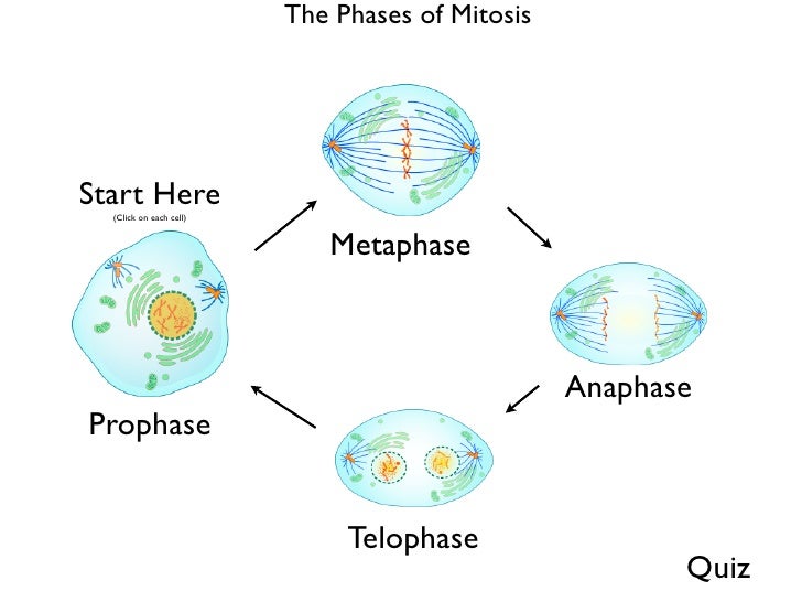
Mitosis (Definition, Diagram & Stages Of Mitosis) - BYJUS List all the stages of mitosis. The stages of Mitosis are: Prophase - The chromosomes shorten and thicken. Metaphase - Chromosomes line up in the middle of the cell. Anaphase - Chromatids break apart at the centromere and move to opposite poles. Telophase - Two nuclei formed after nuclear envelopes reform around each group of chromosomes. Explain Animal Cell With Diagram Labeled - ACTUINDE An animal cell diagram is a great way to learn and understand the many functions of an animal cell. Animal cell Diagram Animal cell size and shape Animal cells come in all kinds of shapes and sizes, ranging in size from a few millimeters to micrometers. We all remember that the human body is quite elaborate and a technique I discovered to are ...
Animal Cell Simple Labeled Diagram - Mitosis Diagram Without Labels For ... The animal cell diagram is widely asked in class 10 and 12 examinations and is beneficial to understand the structure and functions of an animal. The diagram, like the one above, will include labels of the major parts of an animal cell including the cell membrane, nucleus, ribosomes, mitochondria, vesicles, and cytosol.
Label the following diagram of mitosis of an animal cell
PDF Cell Division Animal Cell and Mitosis Key - Scarsdale Public Schools Mitosis has four phases: prophase, metaphase, anaphase, and telophase. Follow the directions. 1. Label the four phases of mitosis in the diagram. 2. Label the spindles and centrioles in one of the phases. 3. Color each chromosome in prophase a different color. Follow each of these chromosomes through mitosis. quizlet.com › 327337663 › mastering-biology-chp-9-hwMastering Biology Chp. 9 HW Flashcards - Quizlet [The key structures involved in mitosis are labeled in this diagram of an animal cell that shows the two sister chromatids of each duplicated chromosome beginning to attach to the mitotic spindle by means of their kinetochores. The centrosomes anchor the mitotic spindle at opposite ends of the cell.] A Well-labelled Diagram Of Animal Cell With Explanation Well-Labelled Diagram of Animal Cell The Cell Organelles are membrane-bound, present within the cells. There are various organelles present within the cell and are classified into three categories based on the presence or absence of membrane. Listed below are the Cell Organelles of an animal cell along with their functions.
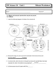
Label the following diagram of mitosis of an animal cell. › cell-division-icse-solutionsICSE Solutions for Class 10 Biology - Cell Division - A Plus ... Dec 04, 2019 · Question 4: (i) Draw a neat labeled diagram to show the metaphase stage of mitosis in an animal cell having ‘6’ chromosome. (ii) How many daughter cells are formed at the end of mitosis and at the end of meiosis? (iii) With reference to cell division explain the following terms: (Chromatid, Centromere, Haploid). (iv) Name the type of cell ... PDF SW Science 10 Unit 1 Mitosis Worksheet - Maplewood Schools 1. Label the following diagram of mitosis of an animal cell. 2. During which stage of a cell's cycle do the replicated chromosomes thicken and become visible? 3. In animal cells, which structure is thought to produce the spindle fibers that help separate the sister chromatids during anaphase? 4. Is this structure found in plant cells? 5. › Cell_BIO › activitiesOnline Onion Root Tips - University of Arizona These regions of growth are good for studying the cell cycle because at any given time, you can find cells that are undergoing mitosis. In order to examine cells in the tip of an onion root, a thin slice of the root is placed onto a microscope slide and stained so the chromosomes will be visible. Solved 39 an The following diagram shows animal cells in | Chegg.com 39 an The following diagram shows animal cells in different phases of the cell cycle and mitosis. Label each cell with the appropriate phase. (Hint: A nucleus with dashed lines indicate that the individual chromosomes are not visible.)
Label the following Diagram of mitosis of an animal cell Step 5: Telophase (Two nuclei produced and cell pinches in the middle) Question # 2: Interphase . Question # 3: Centrioles are the organelles that help in producing spindle fibers. Labelling of individual structures: Structures present at the poles of the cell (Centrioles) Haploid chromatin 1n (Chromatid) Diploid chromatin 2n (Chromosome) › mitosisAnimal Cell Mitosis - CELLS alive Events during Mitosis. Interphase: Cells may appear inactive during this stage, but they are quite the opposite. This is the longest period of the complete cell cycle during which DNA replicates, the centrioles divide, and proteins are actively produced. For a complete description of the events during Interphase, read about the Cell Cycle. DOC Mitosis: Labeled Diagram - West Branch High School The daughter cells are identical to one another and to the original parent cell. In a typical animal cell, mitosis can be divided into stages: Interphase: Gap 1 Phase (growth), Synthesis Phase (copy of DNA), Gap 2 Phase (organelle production) ... Mitosis: Labeled Diagram Author: jana stewart Last modified by: jana stewart Created Date: 1/19 ... quizlet.com › 248407215 › mastering-biology-chapterMastering Biology Chapter 9 Flashcards - Quizlet Through a microscope, you can see a cell plate beginning to develop across the middle of a cell and nuclei forming on either side of the cell plate. This cell is most likely _____. a bacterial cell dividing a plant cell in the process of cytokinesis an animal cell in the process of cytokinesis a plant cell in metaphase
SOLVED:Label the figure below to identify cellular changes ... - Numerade label the figure below to identify cellular changes occurring during mitosis in animal cells telophase and cvtokinesis interphase prophase prophase metaphase anaphase cleavage furrow chromosomes at spindle equator plasma membrane daughter chromosomes chromatin nuclear envelope fragments chromosome_ consisting of two sister chromatids early … PDF Mitosis Worksheet Answers Enchanted Learning LearningAnswers: Mitosis, Animal Cell Label the animal cell mitosis diagram. Answers: Microscope Label the diagram of a microscope. Answers: Neuron Anatomy: Label the Cell Label the axon, dendrites, cell body, nucleus, Schwann's cells, and nodes of Ranvier. Answers: Plant Cell Label the plant cell diagram using the glossary of plant cell terms ... PDF Mitosis Practice - Georgetown ISD In animal cells, which structure is thought to produce the spindle fibers that help ... Based on the drawing, in what stage of mitosis must the cell have been in? Label the following diagram of mitosis of an animal cell. 6. The drawings A-E show stages of mitosis in an plant cell. (a) (b) Which of the drawings A -E shows shorten) (ii) (iii) (iv ... mitosis_1_answers.pdf - SW Science 10 Unit 1 Mitosis... - Course Hero Label the following diagram of mitosis of an animal cell. 2. During which stage of a cell's cycle do the replicated chromosomes thicken and become visible? ______________________ 3. In animal cells, which structure is thought to produce the spindle fibers that help separate the sister chromatids during anaphase? ______________________ 4.
a draw a neat diagram of a plant cell and label the following parts i ...
Labeled Onion Cell Diagram The information can be. Onion Cell Mitosis Background. In a typical animal cell mitosis can be divided into stages. 1 Comparison Of Cross Sectional Images Of Onion Root Tip Cells. Engine Diagram Camry 98 4 marks e The onion epidermal cells are not green in colour because they lack. Onion Cell Mitosis Background.

Pin on Biology Materials-Microorganisms,Protists,Bacteria,Fungi &Viruses
DOC Mitosis Worksheet & Diagram Identification - birdvilleschools.net Cell Cycle, Mitosis, & Cancer Review. Due Date: _____ Using the labeled Cell Cycle diagram to the right do the following: Left column: place the corresponding letter for the correct stage. Right column: Complete the description of the stage in the. A _____: Consists of G1, S and G2 phases in the Cell Cycle. ___ Cytokinesis
Mitosis Worksheet - Mitosis Worksheet Date Name 1 Label the following ...
DOC MITOSIS WORKSHEETS - barajasscience.weebly.com Label the following diagram of mitosis of an animal cell using the Word Bank ( 2. During which stage of a cell's cycle do the replicated chromosomes thicken and become visible? 3. In animal cells, which structure is thought to produce the spindle fibers that help separate the sister chromatids during anaphase? 4.

Mitosis Worksheet - Fill Online, Printable, Fillable, Blank | pdfFiller
› cell › cell-cycle4 Major Phases of the Cell Cycle (With Diagram) The following points highlight the four major phases of the cell cycle. The phases are: 1. G 1 (gap1) phase 2. S (synthesis) phase 3. G 2 (gap 2) phase 4. M (mitosis) phase. Cell Cycle: Phase # 1. G 1 Phase: The G 1 phase is set in immediately after the cell division. It is characterised by a change in the chromosome from the condensed mitotic ...

Biology 2e, The Cell, Cell Reproduction, The Cell Cycle | OpenEd CUNY
Animal Cell Diagram Mitosis Structure : Functions and Diagram Mitosis Diagram showing the different stages of mitosis Mitosis is the phase of the cell cycle where the nucleus of a cell is divided into two nuclei with an equal amount of genetic material in both the daughter nuclei. In animal cells which structure is thought to produce the spindle fibers that help separate the sister chromatids during anaphase.

Apologia Biology | PotterVilla Academics
Solved 1.2 Mitosis and asexual reproduction require one - Chegg Question: 1.2 Mitosis and asexual reproduction require one parent 1.2.1 Mitosis 1. Label the following diagram of mitosis of an animal cell. nghantadilanninn ram 2. During which stage of a cell's cycle do the replicated chromosomes thicken and become visible? 3.
😊 Four phases of mitosis. Diagram of Cell Division by Mitosis. 2019-02-20
i) Draw a well - labelled diagram to show the metaphase stage of ... i) Draw a well - labelled diagram to show the metaphase stage of mitosis in an animal cell having four chromosomes.ii) Mention any two reasons for the population explosion in India.iii) Give biological reasons for the following.1) The pituitary gland is also known as the master gland.2) Gametes have a haploid number of chromosomes. Class 11

Animal Cell Mitosis Label Me! Printout - EnchantedLearning.com
Animal Cell Mitosis Label Me! Printout - Enchanted Learning Anaphase - the phase of mitosis in which the chromosomes begin to separate. Centrioles - paired cylindrical organelles, arranged at right angles to each other, located at the center of a microtubule. Centromeres - a centromere is the constricted region of a nuclear chromosome - microfibers attach to the centromere during mitosis.
Post a Comment for "39 label the following diagram of mitosis of an animal cell"